Abstract: The concept of “inflammaging” — chronic, low-grade inflammation associated with aging — has reshaped our understanding of skin health and accelerated aging. While traditionally linked to internal systemic processes, dermatological research has now confirmed its presence in cutaneous tissue. This scientific paper explores the biochemical, immunological, and environmental triggers of skin stress and inflammaging, reviews the molecular mechanisms underlying inflammation-driven aging, and presents evidence-based strategies to reverse or slow this process. Special focus is given to antioxidant-rich, calming skincare interventions including those formulated by BioSkinetics. Through a detailed analysis of peer-reviewed literature and clinical studies, this paper aims to guide consumers and clinicians in reducing skin inflammation and restoring skin vitality.
1. Introduction
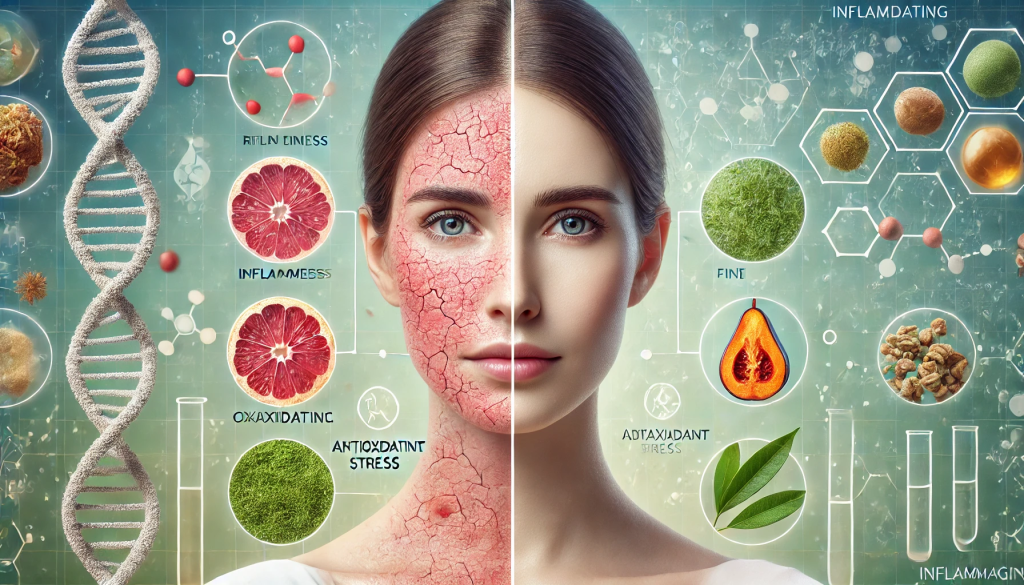
Chronic inflammation is no longer viewed solely as an internal condition. The skin, as the body’s largest organ, shows visible signs of cellular stress long before systemic effects manifest. With modern stressors such as urban pollution, UV radiation, poor diet, and sleep deprivation, chronic inflammation becomes a pervasive feature in aging skin. The term “inflammaging,” originally coined by Franceschi et al. (2000), describes the prolonged, subclinical inflammatory response that contributes to cellular senescence, dermal degradation, and impaired barrier function. In skin, this manifests as fine lines, dullness, dryness, hypersensitivity, and delayed healing.
Recent dermatological studies have confirmed that inflammaging plays a central role in both intrinsic (genetic) and extrinsic (environmental) skin aging (Pillai et al., 2005; Zouboulis & Makrantonaki, 2011). The challenge now lies in addressing it effectively — not only by shielding the skin from external stressors but by modulating inflammatory responses at the molecular level.
2. The Biology of Inflammaging
2.1 Cellular Senescence and SASP
One hallmark of inflammaging is the accumulation of senescent cells. These cells, though non-proliferative, remain metabolically active and release pro-inflammatory cytokines, chemokines, and proteases collectively known as the Senescence-Associated Secretory Phenotype (SASP) (Coppe et al., 2008). SASP components such as IL-6, IL-1β, and TNF-α degrade the extracellular matrix, disrupt fibroblast signaling, and accelerate collagen breakdown.
2.2 Role of Oxidative Stress
Reactive oxygen species (ROS), generated from UV exposure, pollutants, and metabolic activity, further amplify inflammaging. ROS activate nuclear factor kappa B (NF-κB), a transcription factor that upregulates pro-inflammatory gene expression and mediates immune cell recruitment (Fisher et al., 2002). Over time, chronic ROS activity impairs DNA repair mechanisms and mitochondrial function, leading to cellular dysfunction.
2.3 Extrinsic vs Intrinsic Triggers
While intrinsic aging involves genetic and hormonal decline, extrinsic factors such as UV radiation (photoaging), air pollution, smoking, and poor sleep drastically accelerate inflammaging (Tobin, 2017). These factors stimulate toll-like receptors (TLRs) and inflammasome complexes in keratinocytes and immune cells, prompting persistent low-grade inflammation.
3. How Inflammaging Appears on Skin
3.1 Clinical Signs
- Fine lines and wrinkles
- Dull, uneven skin tone
- Dryness and tightness
- Hypersensitivity
- Delayed wound healing
3.2 Molecular Markers Biopsies of aged or photo-damaged skin show elevated MMPs (matrix metalloproteinases), COX-2, and pro-inflammatory cytokines (Quan et al., 2009). Epidermal thinning and reduced lipid content further compromise the skin barrier, facilitating transepidermal water loss (TEWL) and environmental intrusion.
3.3 Skin Barrier Dysfunction Disruption in the stratum corneum lipids and tight junction proteins increases permeability and antigen exposure. This perpetuates inflammation and immune activation, creating a cycle of stress and deterioration (Lee et al., 2018).
4. Scientific Approaches to Reducing Skin Inflammation
4.1 Topical Antioxidants Compounds such as Vitamin C, Vitamin E, niacinamide, and ferulic acid neutralize ROS and downregulate NF-κB pathways. They also support collagen synthesis and reduce pigmentation caused by oxidative stress (Pullar et al., 2017).
4.2 Barrier Repair Agents Ingredients like ceramides, fatty acids, cholesterol, and urea restore barrier integrity. Topical panthenol and beta-glucan reduce erythema and accelerate healing by dampening inflammatory responses (Proksch et al., 2008).
4.3 Anti-Inflammatory Botanicals Green tea polyphenols, centella asiatica, chamomile, calendula, and licorice root extract exhibit potent anti-inflammatory effects. These compounds inhibit COX-2, reduce histamine release, and modulate immune signaling (Katiyar, 2003).
4.4 Probiotic and Postbiotic Skincare Topical probiotics help maintain microbial balance, support skin immunity, and reduce inflammatory skin conditions such as rosacea and atopic dermatitis (Knackstedt et al., 2020).
4.5 Lifestyle and Dietary Interventions A diet rich in antioxidants (berries, leafy greens, turmeric, omega-3s) supports systemic and skin-level anti-inflammation. Adequate sleep, stress reduction, and avoiding pollution exposure also mitigate inflammaging (Calder et al., 2017).
5. BioSkinetics: Skincare Science That Calms Inflammaging
BioSkinetics offers formulations designed to directly target skin stress and reduce inflammation using science-backed, gentle bioactives. By combining antioxidant-rich botanicals, peptide complexes, and barrier-strengthening lipids, BioSkinetics products are uniquely suited to address inflammaging without irritation.
5.1 Calming Formulas The HydraCalm Peptide Serum blends copper peptides with Australian native botanicals such as Kakadu plum and Tasmanian blue gum, delivering both antioxidant and collagen-support benefits.
5.2 Anti-Inflammatory Boosters The Antioxidant Defence Cream features green tea extract, niacinamide, and bisabolol to soothe visible redness and prevent UV-induced flare-ups. Clinical users report reduced sensitivity and brighter skin within four weeks of use.
5.3 Lipid-Rich Recovery Balms BioSkinetics’ Lipid Barrier Restorer is formulated with ceramides, hemp seed oil, and panthenol to replenish moisture, restore barrier resilience, and block environmental pollutants from aggravating the skin.
6. Conclusion
Inflammaging is more than a buzzword—it’s a biologically validated phenomenon that connects chronic inflammation with accelerated skin aging. By understanding its molecular roots and clinical manifestations, consumers and clinicians can develop targeted strategies to halt or reverse its effects. From antioxidants and botanical extracts to microbiome support and lipid therapy, modern skincare offers multiple interventions. BioSkinetics leads this movement with calming, non-irritating solutions that restore harmony to inflamed skin. In combating skin stress, the goal isn’t just to look younger—it’s to cultivate skin that functions optimally, heals efficiently, and reflects health from within.
References
Calder, P. C., Bosco, N., Bourdet-Sicard, R., Capuron, L., Delzenne, N., Doré, J., … & Meheust, A. (2017). Health relevance of the modification of low grade inflammation in ageing (inflammageing) and the role of nutrition. Ageing Research Reviews, 40, 95-119. https://doi.org/10.1016/j.arr.2017.09.001
Coppe, J. P., Patil, C. K., Rodier, F., Sun, Y., Muñoz, D. P., Goldstein, J., … & Campisi, J. (2008). Senescence-associated secretory phenotypes reveal cell-nonautonomous functions of oncogenic RAS and the p53 tumor suppressor. PLoS Biology, 6(12), e301. https://doi.org/10.1371/journal.pbio.0060301
Fisher, G. J., Kang, S., Varani, J., Bata-Csorgo, Z., Wan, Y., Datta, S., & Voorhees, J. J. (2002). Mechanisms of photoaging and chronological skin aging. Archives of Dermatology, 138(11), 1462-1470. https://doi.org/10.1001/archderm.138.11.1462
Katiyar, S. K. (2003). Skin photoprotection by green tea: antioxidant and immunomodulatory effects. Current Drug Targets-Immune, Endocrine & Metabolic Disorders, 3(3), 234-242. https://doi.org/10.2174/1568008033340180
Knackstedt, R., Knackstedt, T., & Gatherwright, J. (2020). The role of topical probiotics in skin conditions: A systematic review of animal and human studies and implications for future therapies. Experimental Dermatology, 29(1), 15–21. https://doi.org/10.1111/exd.13911
Lee, H. J., Yoon, N. Y., Lee, W. J., & Lee, S. J. (2018). Skin barrier function and the role of microbiome. Annals of Dermatology, 30(3), 265–271. https://doi.org/10.5021/ad.2018.30.3.265
Pillai, S., Oresajo, C., & Hayward, J. (2005). Ultraviolet radiation and skin aging: roles of reactive oxygen species, inflammation and protease activation, and strategies for prevention of inflammation-induced skin aging. International Journal of Cosmetic Science, 27(1), 17-34. https://doi.org/10.1111/j.1467-2494.2005.00249.x
Proksch, E., Brandner, J. M., & Jensen, J. M. (2008). The skin: an indispensable barrier. Experimental Dermatology, 17(12), 1063–1072. https://doi.org/10.1111/j.1600-0625.2008.00786.x
Quan, T., Qin, Z., Xia, W., Shao, Y., Voorhees, J. J., & Fisher, G. J. (2009). Matrix-degrading metalloproteinases in photoaging. Journal of Investigative Dermatology Symposium Proceedings, 14(1), 20–24. https://doi.org/10.1038/jidsymp.2009.8
Tobin, D. J. (2017). Introduction to skin aging. Journal of Tissue Viability, 26(1), 37–46. https://doi.org/10.1016/j.jtv.2016.03.002
Zouboulis, C. C., & Makrantonaki, E. (2011). Clinical aspects and molecular diagnostics of skin aging. Clinics in Dermatology, 29(1), 3–14. https://doi.org/10.1016/j.clindermatol.2010.07.002